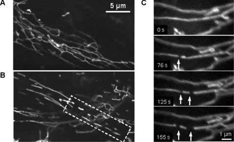
Fig 3.

Fragmentation of mitochondrial network filaments in human pancreatic cells. (A) Continuous mitochondrial network. (B) Partially fragmented mitochondrial network with shorter separated filaments (dashed box). (C) Dynamics of mitochondrial fission. Fast division of long filament into three fragments (arrows) occurred during about 100 sec. (A) and (B): Scale bar, 5 μm. (C): Scale bar, 1 μm. The video file for Fig. 2C is available under ‘Supporting Information’.
