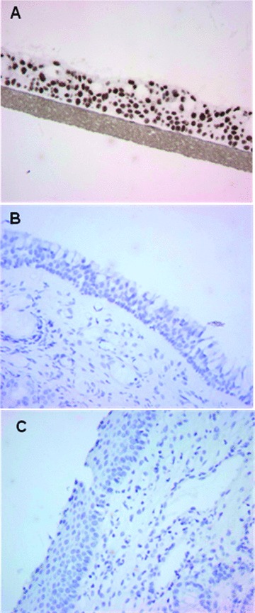
Fig 3.

Immunohistochemical analysis of cyclobutane pyrimidine dimers in nasal epithelial tissues from patients subsequent to treatment with rhinophototherapy. Monoclonal antibodies specific for CPDs were used to visualize DNA damage in the nasal epithelium of patients 2 months after the final exposure to a RPT treatment for seasonal allergies (i.e. 9 treatments over a 3-week period). Tissue sections are shown for RPT-treated patients (C), sham-treated (visible light only) patients (B) and a positive control of UV irradiated 3D reconstructed normal human respiratory epithelium (EpiAirway) (A).
