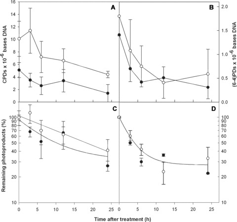
Fig 5.

Nucleotide excision repair kinetics in 3D reconstructed normal human nasal epithelial and epidermal tissues. CPD and (6–4)PD repair are shown in EpiDerm (•–•) and EpiAirway (○–○) artificial tissues. The upper panels show the frequencies of CPDs (A) and (6–4)PDs (B) at increasing times post-irradiation as lesions per megabase DNA. Directly below each panel the frequencies have been normalized to the amount of damage measured at T0 and are expressed as the percentage of CPDs (C) and (6–4)PDs (D) remaining at increasing times post-irradiation with a single sublethal dose of UVB. Exponential decay curves for each data set are shown. Standard errors of the mean were calculated from standard deviations using a total of 8 data points from duplicate tissues.
