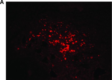
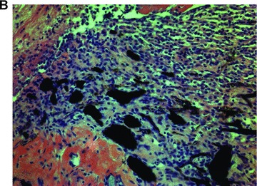
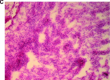
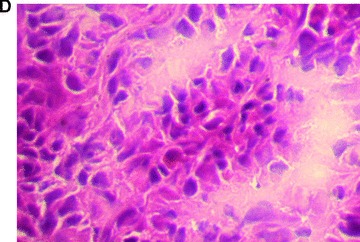
Fig 3.


Morphology of engrafted foetal liver cells (FLC) in the explanted neo-tissue at day 14 after transplantation. (A) Fluorescence microscopy revealed the pkh+ transplanted FLC within the neo-tissues (10 × 4). (B) Haematoxylin and eosin staining (10 × 4) of a serial section showed the engraftment of cell groups in close relationship to the neo-capillarization (seen by India ink staining). (C) Haematoxylin and eosin staining 10 × 10. Higher magnification of cell groups demonstrated that polygonal shaped cells with prominent nuclei formed a neo-tissue within the fibrin matrix in vicinity to the central AV-loop vessel. (D) Haematoxylin and eosin staining, detail of 3C (10 × 20). Foetal hepatocytes formed a three-dimensional tissue permitting high cell density and cell-to-cell contacts within the matrix in the neo-tissue.
