
Fig 1.
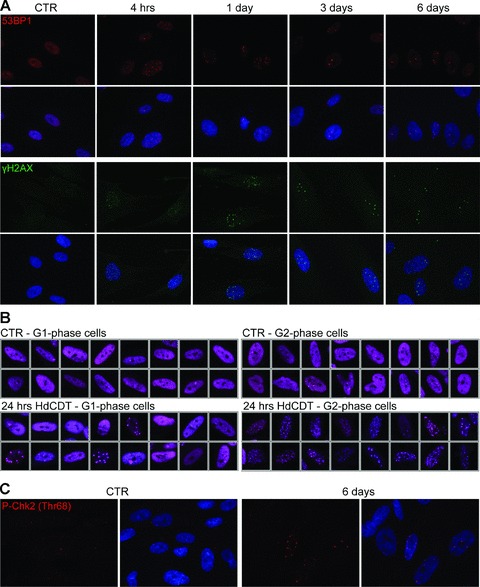
Persistent DNA damage foci in HdCDT-intoxicated cells. (A) BJ fibroblasts were fixed and stained with anti-53BP1 (red) or anti-γH2AX (green) antibody and DAPI (blue) at different times after HdCDT intoxication. (B) Control IMR-90 cells and IMR-90 cells exposed to HdCDT for 24 hrs were fixed and stained with anti-53BP1 (purple) and DAPI. Depending on an amount of incorporated DAPI dye into the DNA, the cells were separated into the G1- and G2-phase population. (C) Six days after the intoxication with HdCDT, IMR-90 fibroblasts were fixed and stained with anti-P-Chk2 (Thr68) (red) and DAPI (blue). Untreated cells were used as negative control (CTR).