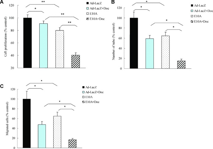
Fig 1.

The effects of E10A and docetaxel on biological function of HUVECs. (A) Proliferation assay. HUVECs were treated with CM from infected PC-3 cells or/and 0.1 nM docetaxel, and cell proliferation was evaluated after 72 hrs. Columns, cell proliferation normalized against Ad-LacZ control (mean ± S.D.). (B) Tube formation assay. HUVECs were treated with CM or/and 0.1 nM docetaxel. Cells were plated on 96-well plates with Matrigel. After 16 hrs of incubation, images of tube formation were captured, and tube formation was scored in one ×50 microscopic field. The number of tube formation was quantitated, and the data are presented as the mean ± S.D. per field (×50) and were normalized to Ad-LacZ-treated control (n= 5). *P < 0.001; (C) Migration assay. HUVECs were treated CM or/and 0.1 nM docetaxel and pipetted into inserts of Matrigel-coated transwells. Addition of chemoattractant, HUVECs complete medium containing 50 ng/ml VEGF, to the lower well of Boyden chamber stimulated endothelial migration to the underside of the transwell membrane. After 6 hrs of incubation, migrated cells were stained by DAPI. The number of cells that migrated was counted by microscopy, and the data are presented as the mean ± S.D. per field (×100) and were normalized to Ad-LacZ-treated control (n= 6), *P < 0.001.
