
Fig 3.
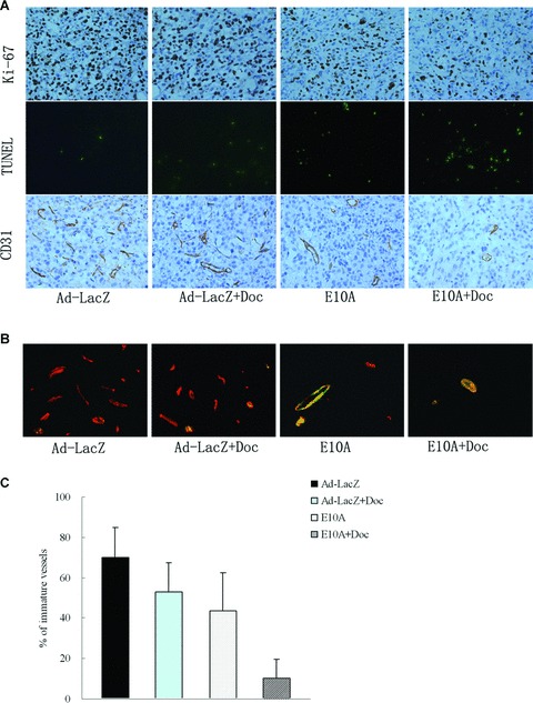
Immunohistochemical analyses PC-3 s.c. tumours in different treatment groups. (A) The sections were immunostained for expression of Ki-67 (cell proliferation), TUNEL (cell apoptosis) and CD31 (endothelial cells). Representative images were shown (×200). (B) Vessels were stained with rabbit anti-CD31 (red) and anti-mouse α-smooth muscle actin (green) to identify whether vessels are mature. Representative images of vessels positive for CD31 (endothelium cells) and α-smooth muscle actin (pericytes) were shown (×400). (C) Determination of immature vessels. Ten independent fields at ×400 were used in morphometric analysis to determine whether vessel is mature. Columns, mean ± S.D.