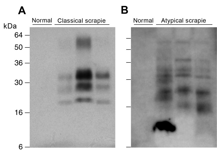
Figure 1.
Detection of disease-related prion protein (PrPSc) in brains of sheep with field cases of classical and atypical scrapie, Germany. Both panels show immunoblots of proteinase K–digested brain homogenate analyzed by enhanced chemiluminescence with monoclonal antibody ICSM 35 against PrP. Samples in panel A are from 10 μL 10% (w/v) brain homogenate after direct digestion with protease. Samples in panel B are derived after processing 200 μL10% (w/v) brain homogenate as described in Materials and Methods. A) From left, normal sheep brain compared to classical scrapie sheep brain samples FLI 1/06, FLI 83/04 and FLI 107/04. B) From left, normal sheep brain compared to atypical sheep scrapie brain samples FLI S7/06, FLI 14/06 and FLI 26/06.

