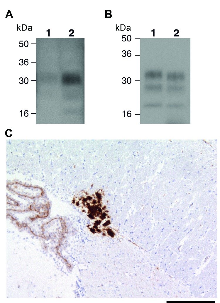
Figure 3.
Ovine bovine spongiform encephalopathy (BSE) prion transmission to a 129MM Tg35c mouse. Panel A shows immunoblot detection of disease-related prion protein (PrPSc) in 10 μL of proteinase K (PK)–digested 10% (w/v) brain homogenates from ovine BSE (SE 1929/0877) (lane 1) and secondary passage ovine BSE (SE1945/0032) (lane 2) using monoclonal antibody ICSM35 against prion protein (PrP). Panel B shows type 4 PrPSc in 1 μL of PK-digested 10% (w/v) vCJD brain homogenate (lane 1) in comparison to PrPSc in 20 μL of PK-digested 10% (w/v) brain homogenate from a 129MMTg35c mouse with subclinical prion infection that was culled 661 days after inoculation with secondary passage ovine BSE inoculum SE1945/0032 (lane 2). Panel C shows abnormal PrP immunoreactivity stained with monoclonal antibody ICSM35 against PrP in the corpus callosum of the ovine BSE–affected 129MM Tg35c mouse brain. Scale bar indicates 165 μm.

