
Figure 1.
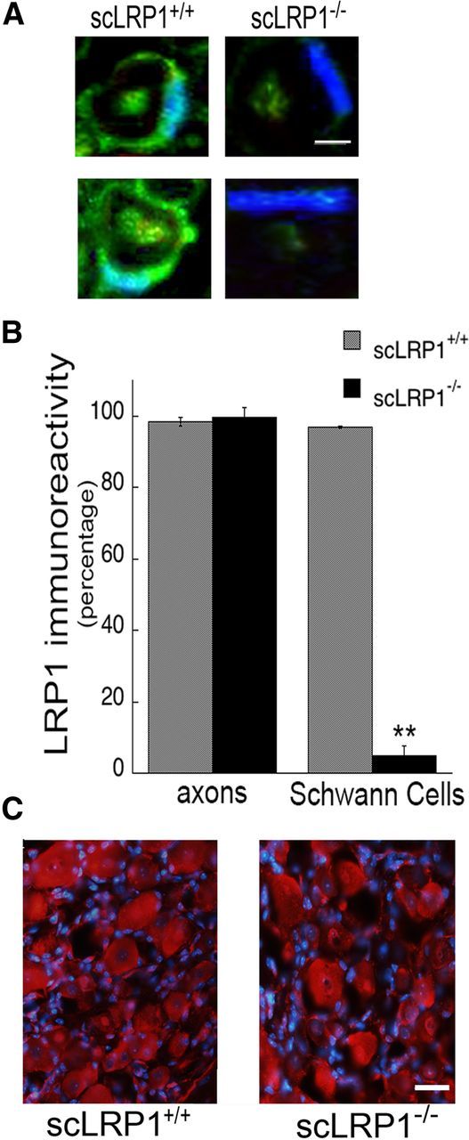
LRP1 inactivation in Schwann cells. A, Double-label immunofluorescence microscopy of LRP1 (green) in an adult myelinated sciatic nerve fiber. Nuclei are identified with DAPI (blue). Note some residual LRP1 immunoreactivity in axoplasm of scLRP1−/− nerves not associated with Schwann cells. Images represent n = 4 mice/group. Scale bar, 4 μm. B, Quantification of LRP1 immunoreactive Schwann cells and axons in uninjured sciatic nerves. Immunoreactive cells were counted in 4 separate sections from each mouse (*p < 0.01; n = 4 mice/group). C, Immunofluorescence microscopy of LRP1 (red) in DRGs. Note similar intensity and distribution of immunoreactivity in DRG neuron cell bodies. Nuclei are identified with DAPI (blue). Scale bar, 45 μm.