
Figure 5.
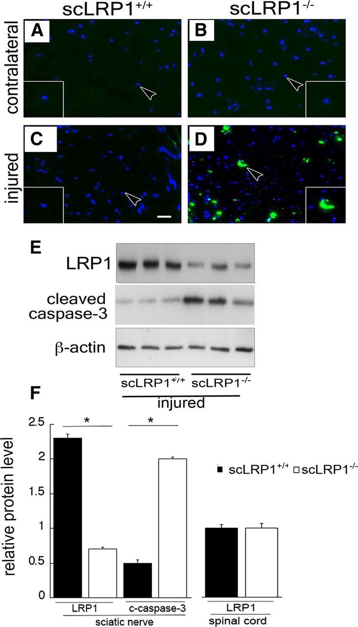
Schwann cell viability is compromised in scLRP1−/− mice. A–D, Sections of crush-injured and control, contralateral sciatic nerves were analyzed by TUNEL-staining (green). Nuclei are stained blue with DAPI. Images are representative of those observed with 3 mice/cohort. Scale bar, 15 μm. E, Immunoblot analysis of LRP1 (85 kDa) and cleaved caspase-3 in sciatic nerve distal to a crush injury site. Nerves were isolated 1 d after injury. Equal amounts of nerve protein (30 μg) were loaded in each lane and subjected to SDS-PAGE. β-Actin was measured as a loading control. Each lane represents an individual animal (n = 3/group). F, Quantification of LRP1 in sciatic nerve and cleaved caspase-3 by densitometry. Quantification of LRP1 in spinal cord by densitometry. Data are expressed as mean ± SEM (*p < 0.05).