
Figure 9.
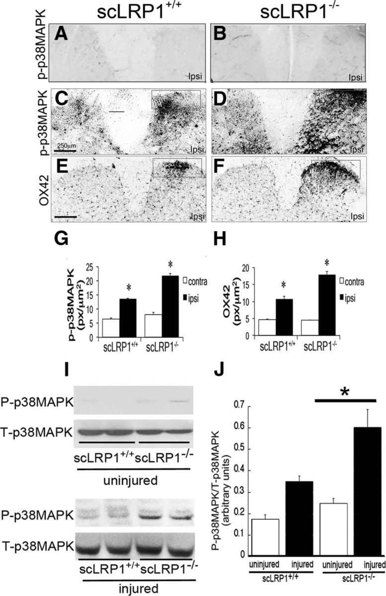
Sustained pain-related behavior in scLRP1−/− mice is associated with phosphorylation of p38 MAPK (P-p38MAPK) and activation of microglia in the spinal dorsal horn. A–D, Immunofluorescence for P-p38MAPK in the spinal dorsal horn of mice in uninjured (A, B) and injured (C, D) 7 d after PNL. E, F, Immunofluorescence of cd11b/OX-42 in the spinal dorsal horn after 7 d of PNL injury. Images are representative of those obtained in studies with 3–4 mice per cohort. Each pair of images shown in A–F (A and B–D, E and F) is matched for exposure. Scale bar, 400 μm. G, H, Quantification by densitometry of P-p38MAPK and OX-42 in the ipsilateral and contralateral spinal dorsal horn. Signal intensity in the ipsilateral horn was significantly increased in scLRP1−/− mice compared with scLRP1+/+ mice (*p < 0.05, n = 4/group). Signal intensity also was significantly increased in the ipsilateral compared with the contralateral horn for each biomarker (*p < 0.05). I, Immunoblot analysis of P-p38MAPK in spinal cord dorsal horn isolated 7 d after PNL. Equal amounts of protein (50 μg) were loaded in each lane and subjected to SDS-PAGE. Total p38 MAPK was determined as a loading control. J, Quantification of P-p38MAPK to T-p38MAPK ratio by densitometry in uninjured and injured spinal cord (n = 3–4 mice) *p < 0.05 comparing injury in each genotype.