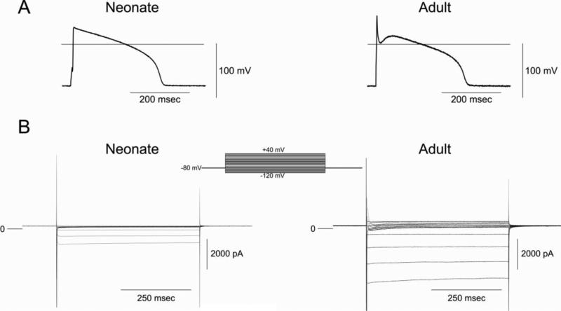
Figure 3.
Panel A: Representative action potentials recorded from a 2 week-old neonate and adult ventricular myocytes. BCL= 2 s. Panel B: Comparison of membrane currents recorded from a neonate and adult venticular cell. The myocytes were held at −80 mV and membrane currents were elicited by stepping the voltage to membrane potentials between −110 and +50 mV.

