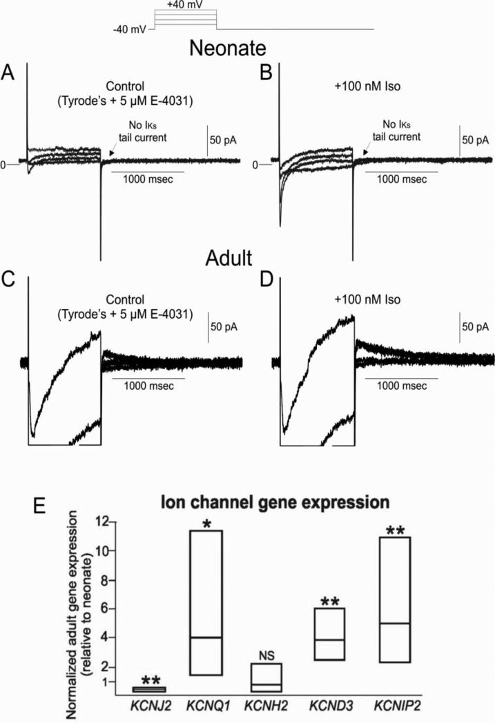
Figure 7.
Panel A: Representative IKs currents from 2 week-old neonate ventricular myocyte. E-4031 (5 μM) was present to eliminate IKr. IKs tail currents were not observed at any potential studied. Panel B: Protocol repeated in the presence of 100 nM isoproterenol. ICa was greatly augmented, although IKs was still absent. Panel C: Representative IKs currents from an adult ventricular myocyte. E-4031 (5 μM) was present to eliminate IKr. IKs tail currents were readily observed in adults using the same protocol. Panel D: IKs was augmented in the presence of 100 nM isoproterenol. Panel E: Ion channel gene expression in adult ventricular tissue (relative to neonatal). Data are presented as box plots with the mean together with upper and lower confidence levels (95%). KCNQ1, KCND3 and KCNIP2 are higher in adult compared with neonate, while KCNH2 is unchanged. KCNJ2 is less in the adult ventricle compared with neonate.

