Figure 8.
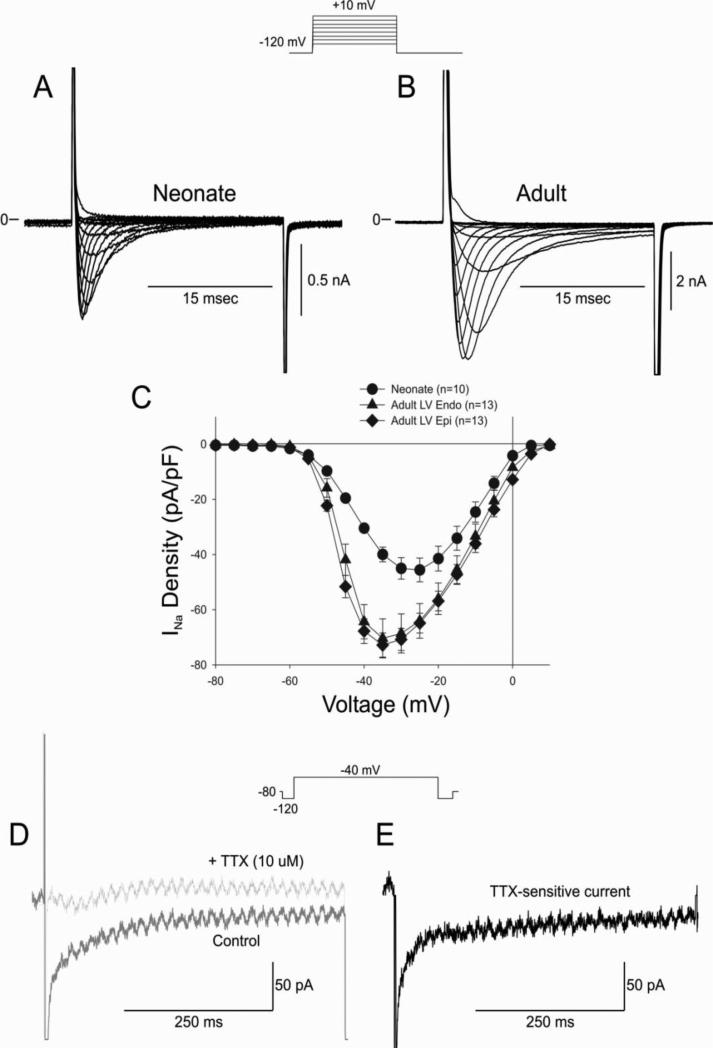
Representative whole cell INa recordings from a 2 week neonate ventricular myocyte (Panel A) and adult ventricular myocyte (Panel B). Current recordings were obtained at test potentials between −80 and 10 mV in 5 mV increments from a holding potential of −120 mV. Panel C: I-V relation for 2 week neonate showing a significantly smaller INa compared to adult LV Epi or Endo. Panel D: Representative late INa recorded during a train of 5 pulses in control solution and after application of TTX (10 μM). Panel E: Subtraction of the traces shown in Panel D yielded the TTX-sensitive late INa.

