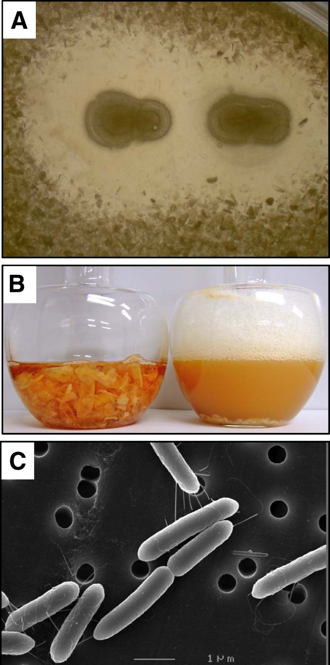
Fig 1.
Chitin degradation and cell morphology of Paenibacillus sp. strain FPU-7. (A) Colonies of strain FPU-7 on an α-chitin powder plate. Cells of strain FPU-7 were smeared on a 1.0% (wt/vol) α-chitin powder plate, and the plate was incubated at 30°C for 1 week. (B) Strain FPU-7 was grown at 30°C for 5 days with shaking, in bonito extract medium containing 5.0% (wt/vol) crab shell chitin flakes. The solid matter (chitin flakes) in the left flask disappeared after 5 days of incubation (right flask). (C) Cell morphology of strain FPU-7 (stationary phase) observed by SEM. Bar, 1 μm.

