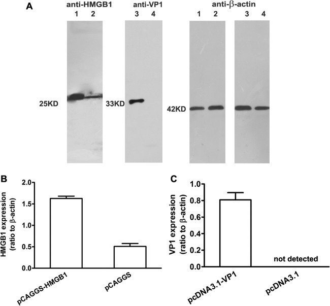
Fig 1.
Expression of VP1 and HMGB1 plasmids in vitro. 293T cells were transfected with various plasmids using Lipofectamine for 48 h, and then cell lysates were subjected to Western blot analysis using anti-VP1, anti-HMGB1, or anti-β-actin antibody. (A) In vitro expression of pHMGB1 and pVP1 plasmids. Lane 1, 293T cells transfected with pCAGGS-HMGB1-HA; lane 2, 293T cells transfected with pCAGGS-HA; lane 3, 293T cells transfected with pcDNA3.1-VP1; lane 4, 293T cells transfected with pcDNA3.1. Molecular mass markers are also shown. (B and C) Quantification of pHMGB1 (B) and pVP1 (C) expression by densitometry. Experiments were repeated three times, with similar results. All of the experimental data were pooled and are presented together.

