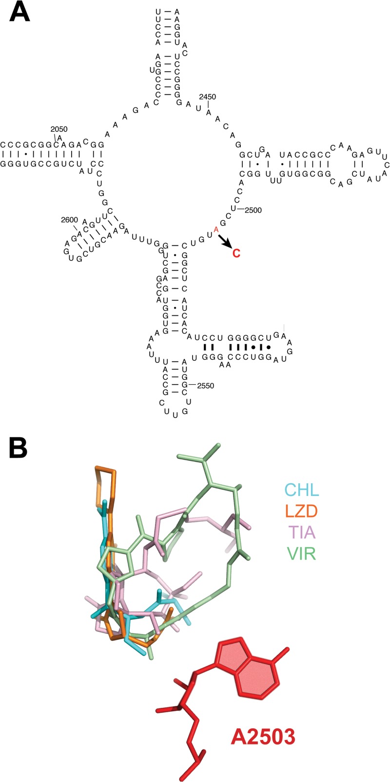
Fig 3.
Mutation, selected in the SQ110DTC strain, conferring resistance to the inhibitor present in extract of strain G006. (A) Position of the mutation within the central loop of domain V of 23S rRNA that participates in formation of the active site of the PTC. (B) Placement of A2503 (red color) in the PTC relative to the binding sites of antibiotics that inhibit peptide bond formation. The aligned structures of the ribosome antibiotic complexes were taken from the DARC server (http://darcsite.genzentrum.lmu.de/darc/) (56). The following antibiotics are shown: virginiamycin M1 (streptogramin A) (VIR), PDB 1YIT; linezolid (LZD), PDB 3CPW; tiamulin (TIA), PDB 3G4S; and chloramphenicol (CHL), PDB 3OFC.

