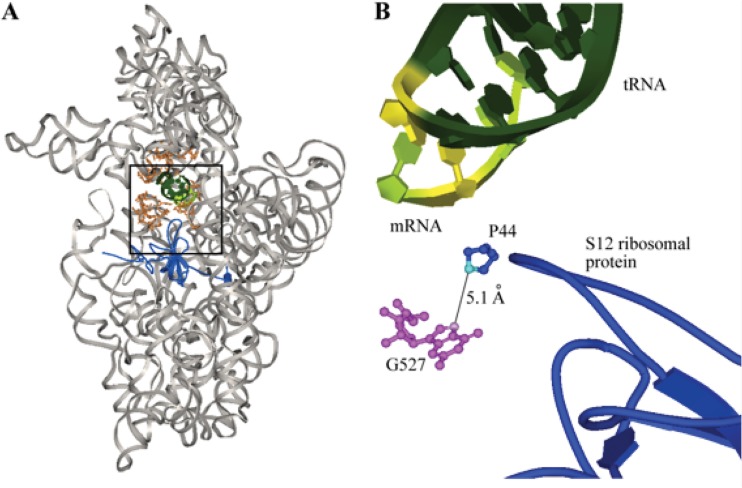
Fig 1.
(A) Crystal structure of the 30S ribosomal subunit of Thermus thermophilus complexed with mRNA and cognate tRNA in the A site (Protein Data Bank [PDB] accession no. 1IBM). 16S rRNA is shown in gray, the A site is in orange, the S12 ribosomal protein is in blue, tRNA is in dark green, and mRNA is in light green. The black box highlights the region shown in panel B. (B) The N7 atom (pink) of G527 (magenta balls and sticks), which corresponds to G518 in M. tuberculosis, is approximately 5 Å; from the C4 atom (turquoise) of proline 44 (blue balls and sticks) of the S12 ribosomal protein (blue tube). The S12 ribosomal protein interacts with the wobble position (highlighted in yellow) of the mRNA:tRNA codon:anticodon helix (dark and light green) (9).

