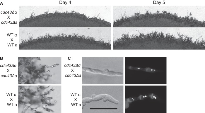
Fig 6.

Role of Ggtase-I in mating. (A) The indicated strains were coincubated in the dark on MS medium. The same location on each plate was imaged for 4 and 5 days. (B) Basidia and spore structures were imaged from matings shown in panel A. (C) Mating plugs were cut, permeabilized, and stained with calcofluor white to visualize the cell wall and with SYTOX green to visualize nuclei. Scale bar, 20 μm.
