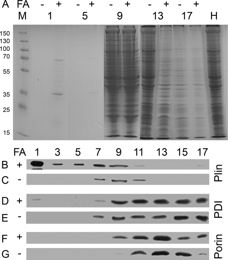
Fig 2.

Purified lipid droplets contain a very limited set of proteins. (A) Cell homogenates from GFP-Plin-expressing untreated cells (−) or cells supplied with fatty acid (FA; +) were resolved on sucrose gradients by ultracentrifugation. Equal volumes taken from the gradient were loaded onto protein gels side by side, separated by electrophoresis, and stained by Coomassie blue. Although all 17 fractions of the gradient were analyzed on a total of three gels, only every fourth fraction (as numbered) was cut out and assembled into this panel. The assembly is flanked by a size marker (M; values in kDa) on the left and the total homogenate (H) on the right. (B to G) For Western blot analysis of the samples, every second fraction (as numbered) was taken, and GFP-perilipin (B and C), the protein disulfide isomerase (PDI) (D and E), or mitochondrial porin (F and G) was detected by the corresponding monoclonal antibody.
