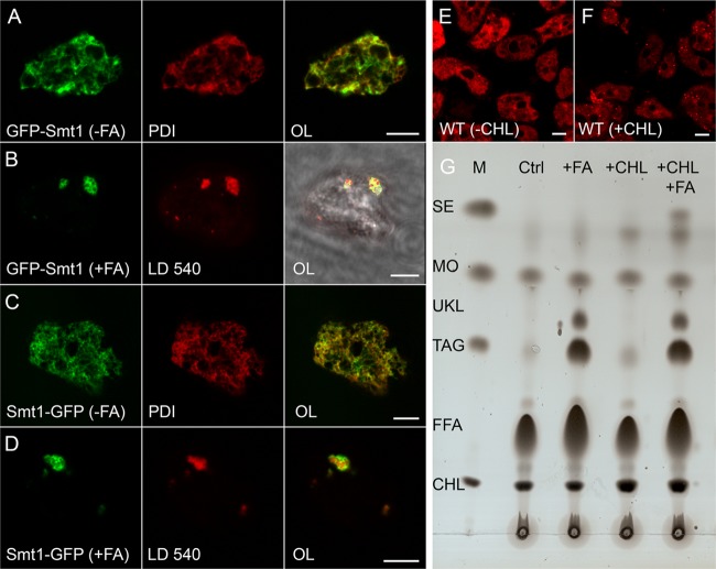
Fig 3.
Dictyostelium lipid droplets contain steryl esters. (A to D) Confocal images from fixed cells expressing steryl methyltransferase 1 (Smt1) tagged with GFP (green channel) at the N-terminal end (A and B) or at its C terminus (C and D) and incubated with (B and D) or without (A and C) fatty acid (FA). The endoplasmic reticulum was revealed by virtue of an antibody directed against PDI that appears red in panels A and C. Alternatively, lipid droplets were stained by LD540 (red in B and D). The overlaid images (OL) appear in the third column (scale bar, 5 μm), where for row B the image from transmitted light is also shown to demonstrate the outline of the otherwise barely visible cell. (E and F) Optical sections through living wild-type (WT) cells stained with LD540 (red) to reveal lipid droplets (dots in panel F) in cells fed with cholesterol (+CHL) for 3 h. In control cells (−CHL) the dye associates nonspecifically with organelle membranes such as the nuclear envelope and the closely associated Golgi apparatus (E). Scale bar, 5 μm. (G) Thin-layer chromatography of lipid samples extracted from wild-type cells grown in axenic medium without further additives (Ctrl), with 200 μM palmitic acid added (+FA), with 100 μM cholesterol (+CHL) added, or with both (+CHL +FA). Substances in the marker lane (M) are labeled as in Fig. 1D. Here, only steryl esters (SE) are relevant. An unknown lipid species (UKL) is further discussed in the text.

