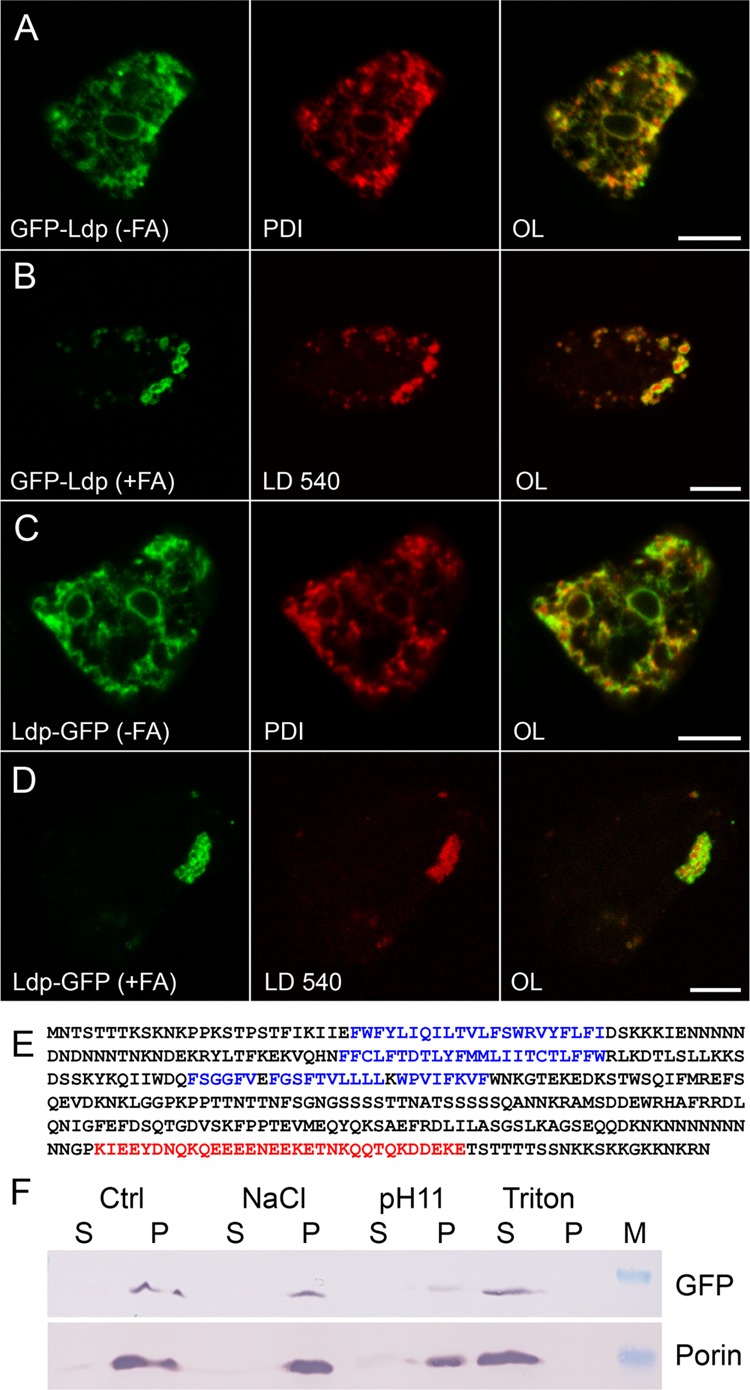
Fig 4.

The novel protein Ldp moves from the ER to lipid droplets. (A to D) Single confocal planes through fixed cells expressing Ldp fused to GFP (green channel) at the N-terminal end (A and B) or carrying the GFP tag at the C terminus (C and D) and incubated in control medium (A and C) or in the presence of palmitic acid (B and D). The endoplasmic reticulum was revealed by immunofluorescence staining with anti-PDI (A and C), whereas lipid droplets were revealed by LD540 (B and D). The overlaid images (OL) show red and green channels. Scale bar, 5 μm. (E) Amino acid sequence of Ldp displayed in one-letter code (60 residues per line). Possible transmembrane segments are shown in blue; a region with coiled-coil character is printed in red. For other features of the protein, see the text. (F) Western blot of supernatant (S) or pellet (P) samples from separating a homogenate derived from Ldp-GFP-expressing cells incubated with homogenization buffer alone (Ctrl), 1 M NaCl, or Na2CO3 at pH 11 (pH 11) to liberate weakly or tightly associated membrane proteins, respectively. Alternatively, Triton X-100 was used to extract transmembrane proteins. The upper band is GFP-tagged LdpA detected by antibody 264−449−2; the lower band represents porin, a protein spanning the outer mitochondrial membrane.
