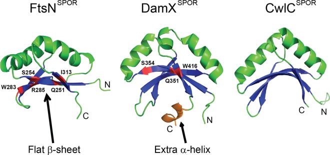
Fig 1.

SPOR domains have similar structures. The 4-stranded β-sheet and 2 α-helices common to all SPOR domains are shown in blue and green, respectively. For clarity, turns and coils are also illustrated in green, while the C-terminal helix unique to DamXSPOR is shown in orange. Residues important for septal localization are highlighted in red. Coordinates for these domains were obtained from the Protein Data Bank (PDB entries 1x60, 1UTA, and 2LFV) and rendered using PyMOL.
