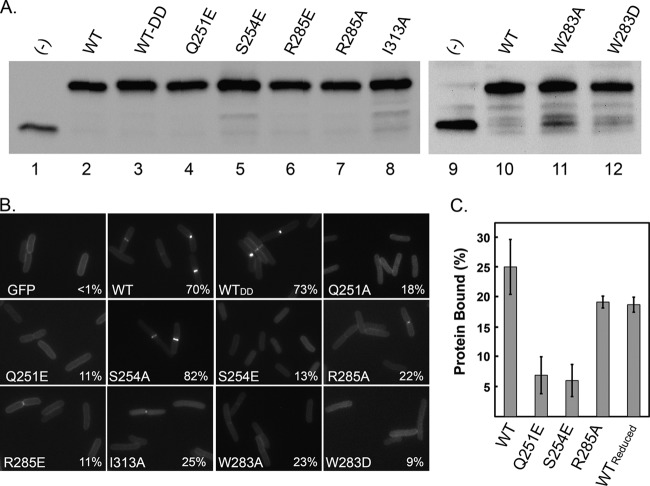
Fig 3.
Localization-defective mutants of FtsNSPOR. (A) Western blot with anti-GFP antibody demonstrating that wild-type (WT) and mutant TTGFP-FtsNSPOR proteins were produced at similar levels. (−), control strain producing TTGFP not fused to anything. (B) Fluorescence micrographs of cells producing the indicated TTGFP-FtsNSPOR fusion proteins. Numbers in the corners are percentages of cells scored as exhibiting septal localization. See Table S3 in the supplemental material for more detailed quantitative data on localization frequencies. (C) Results of PG binding assay. “Reduced” refers to the presence of 1 mM DTT. Bars represent the means and standard deviations for at least three independent experiments.

