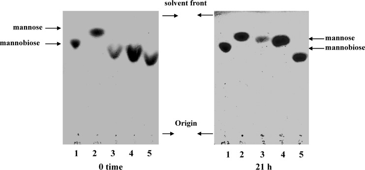
Fig 1.
Hydrolysis of α-mannobioses by sonicated cell extracts of P. gingivalis detected by TLC on Keiselgel F254 HPTLC plates in n-butanol–ethanol–water (5:5:3, by volume). α-Mannosidase assays using α-1→2-, α-1→3-, and α-1→6-linked mannobioses were performed as described in Materials and Methods, and the assay mixtures contained 125 μg of disaccharide in 30 μl of 0.125 M sodium acetate buffer, pH 6.0, with 1.25 mM Ca2+. Ten-microliter aliquots were withdrawn and stored at 4°C, and this served as the zero time point. Ten microliters of sonicated cell extracts of P. gingivalis was added to the reaction mixture and incubated at 37°C for 20 h. Aliquots (10 μl) were withdrawn and centrifuged in an Eppendorf microcentrifuge. The supernatant was spotted onto Keiselgel F254 HPTLC glass-coated plates, and TLC was performed in a tank equilibrated for 1 h in n-butanol–ethanol–water (50:50:30, by volume). The plates were dried in air, rechromatographed in the same solvent system, dried in air, sprayed with 3% sulfuric acid in methanol, air dried, and placed in an oven at 85°C to develop the spots. Lanes: 1, α-1→2 mannobiose; 2, mannose; 3, α-1→2 mannobiose plus P. gingivalis cell extract; 4, α-1→3 mannobiose plus P. gingivalis extract; 5, α-1→6 mannobiose plus P. gingivalis extract.

