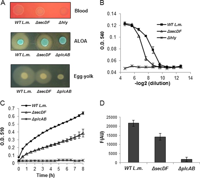
Fig 4.
ΔsecDF mutant exhibits low activity of LLO, PlcA, and PlcB. (A) Activity assays of LLO, PlcA, and PlcB in WT L. monocytogenes and the ΔsecDF mutant grown on blood, ALOA, and egg yolk indicative agar plates, respectively. The halo (clear or opaque) zone around the bacterial patch is proportional to the LLO, PlcA, and PlcB activity. The pictures shown are representative of 3 independent experiments (n = 3). (B) LLO activity hemolysis assay based on lysis of sheep red blood cells. LLO activity was measured in the supernatants of WT L. monocytogenes and the ΔsecDF mutant grown overnight in LB-Glu-1P media. The Δhly mutant was used as a control. The data are representative of three biological repeats (n = 3). The error bars represent standard deviations from triplicate trials. (C) PI-PLC (PlcA) activity assay, measuring reaction turbidity (adapted from Geoffroy et al. [46]). PI-PLC activity was measured in supernatants of WT L. monocytogenes and ΔsecDF mutant grown overnight in LB-Glu-1P media. The ΔplcAB mutant was used as a control. The data are representative of three biological repeats (n = 3). Error bars represent the standard deviations from triplicate trials. (D) A measurement of PC-PLC (PlcB) activity using the EnzChek direct phospholipase C assay kit (Molecular Probes). PC-PLC activity was measured in supernatants of WT L. monocytogenes and the ΔsecDF mutant grown overnight in LB-Glu-1P media. The ΔplcAB mutant was used as a control. The data are representative of three biological repeats (n = 3). Error bars represent the standard deviations from triplicate trials.

