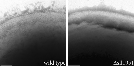
Fig 2.

Phosphotungstic acid negative-stained images of cells of the wild-type (left) and Δsll1951 (right) strains. A honeycomb-like structure of the surface is visible at the periphery of the wild-type strain; it shifts out of the focal plane toward the bottom of the image. The S-layer is absent in the Δsll1951 cell. Bar, 100 nm.
