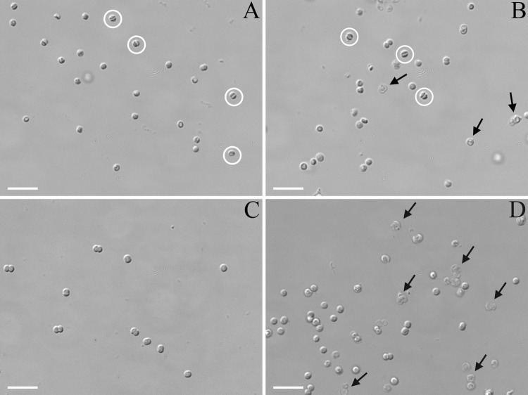
Fig 3.
DIC light microscopy images of Synechocystis wild-type (A and C) or Δsll1951 (B and D) cells after lysozyme treatment, followed by either hyperosmotic shock (A and B) or hypo-osmotic shock (C and D). Control cells (not lysozyme treated) resembled the cells shown in panel C. Water efflux caused significant cell shrinkage (exemplified by cells with white circles) in many cells of both strains and some cell lysis in the Δsll1951 strain (exemplified in cells indicated by arrows). The Δsll1951 cells were significantly more susceptible to the hypo-osmotic effects than the wild type; this shock led to significant lysis and the appearance of ghost-like cells (exemplified in cells indicated by arrows). Bar, 5 μm.

