Fig 1.
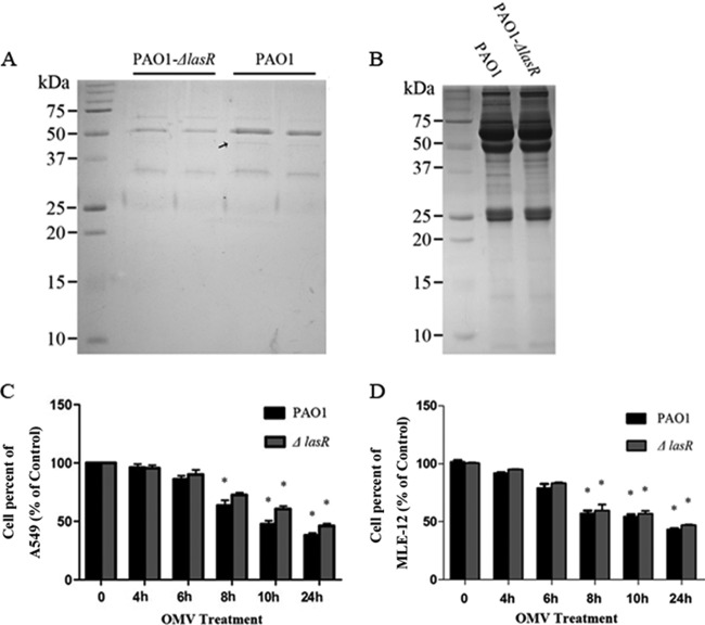
P. aeruginosa PAO1- and PAO1-ΔlasR-generated OMVs elicited time-dependent cytotoxicity in lung epithelial cells. (A and B) Fractions sequentially removed from the top of each gradient were analyzed with Coomassie-stained SDS-PAGE gels. (A) Low-density fractions; (B) high-density fractions. The black arrow indicates the different band between the components of PAO1- and PAO-ΔlasR-generated OMVs. (C and D) Cytotoxicity was determined in A549 cells (C) and MLE-12 cells (D) by an MTT assay. Ten microliters of purified vesicles (0.25 mg/ml) from PAO1 or PAO1-ΔlasR was added to each well and incubated for the indicated time points. Cells treated with an equal amount of PBS were used as a control. Data are presented as means ± SEM, and each column is compared with the 0-h time point (*, P < 0.05, determined by one-way ANOVA followed by a Tukey-Kramer post hoc test).

