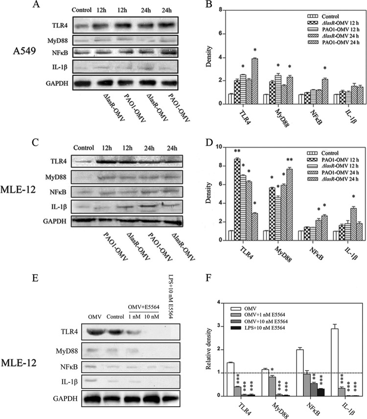
Fig 2.
P. aeruginosa OMVs trigger intracellular inflammatory responses via the TLR4 signaling pathway. (A to D) Released P. aeruginosa OMVs at a concentration of 0.25 mg/ml increased the expression levels of TLR4 and immunogenic proteins MyD88 and NF-κB, finally causing the release of proinflammatory factors in A549 cells (A and B) and MLE-12 cells (C and D), as determined by Western blotting. Cells treated with the same amount of PBS were used as a control. (E and F) Production of TLR4 immunogenically related proteins was significantly impaired when TLR4 was inhibited by E5564 at a concentration of 10 nM. Cells treated with the same amount of PBS were used as a control group. Cells treated with LPS at a concentration of 100 ng/ml and 10 nM E5564 were used as a control for E5564. Representative gels are depicted with a graph that shows the means ± SEM from 3 experiments in which protein levels were normalized to GAPDH levels (B and D) and then to levels of a PBS-treated control (F). Levels shown in each column were significantly decreased compared with the levels of the control (B and D) or natural OMVs (F) (*, P < 0.05; **, P < 0.001 [determined by one-way ANOVA followed by a Tukey-Kramer post hoc test]).

