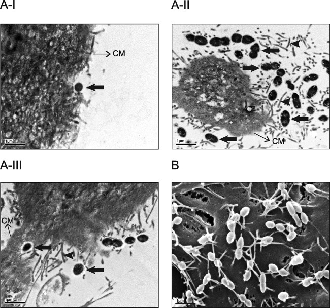
Fig 4.
TEM and SEM showing interactions between S. suis serotype 2 and NTPr cells. (A-I) TEM of S. suis serotype 2 strain 31533 infection of virus-free (control) NPTr cells showing very few cocci at the cell surface. (A-II and A-III) TEM of S. suis strain 31533 infection of swH1N1-preinfected NPTr cells showing high numbers of cocci interacting with epithelial cells (A-II) and intracellular bacteria (A-III). Scale bar, 1 μm. Original magnification, ×5,000. (B) SEM of S. suis serotype 2 strain 31533 infection of swH1N1-preinfected NPTr cells showing high numbers of cocci intimately interacting with cell cilia. Scale bar, 1 μm. Original magnification, ×10,000. No bacteria could be found in the observed SEM fields of control NPTr cells infected with S. suis strain 31533 only (data not shown). Black arrows show bacterial cells, and arrowheads show cilia. CM, cell membrane.

