
Fig 7.
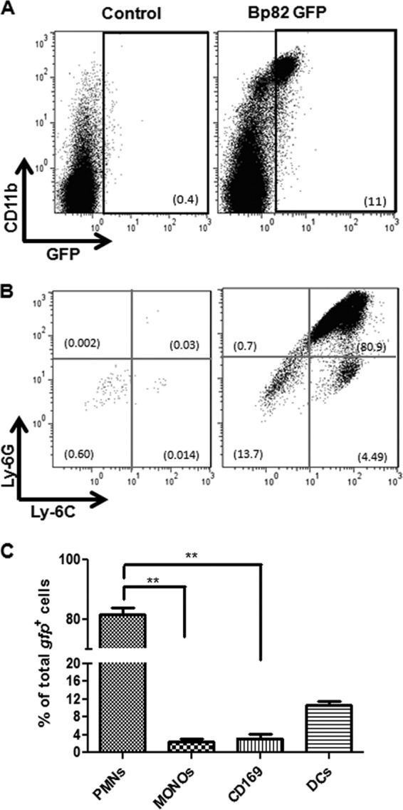
Lymph node cellular response to Bp82 vaccination. BALB/c mice (n = 5 per group) were injected in one rear limb footpad with 1.2 × 10⁸ CFU of Bp82-gfp. Ten hours after inoculation, cells from the ipsilateral popliteal LN were collected and immunostained. Cells from the contralateral popliteal LN served as the control. (A) The appearance of GFP+ CD11b+ cells in the draining LN was determined by flow cytometry at 10 h after injection. (B) The GFP+ cells were further subdivided into polymorphonuclear leukocytes (PMN) (CD11b+ Ly6G+ Ly6C−) and monocytes (CD11b+ Ly6G− Ly6C+). (C) The distribution of Bp82-gfp bacteria in relevant LN APC populations, including neutrophils (PMN), dendritic cells (DC), CD169+ subcapsular macrophages, and monocytes, was calculated. Data are representative of two independent experiments. Statistical differences between Bp82-injected and uninjected contralateral LN were determined by Student's two-tailed t test. **, P < 0.05. For comparisons between 4 groups (C), ANOVA was used, followed by the Tukey post hoc test.