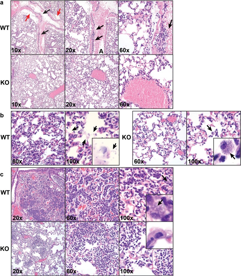
Fig 2.
Histopathological analysis of A. baumannii-induced immune response in Fus1+/+ (WT) and Fus1−/− (KO) lungs at different time points after infection. (a) 4 hpi, H&E staining. The upper row shows different magnifications of Fus1−/− lungs distinguished by the presence of neutrophils in the interstitium with minimal leukocyte margination observed in blood vessels. In contrast, the bottom row illustrates the late response to bacterial challenge in Fus1+/+ lungs characterized by margination of leukocytes (black arrows) and perivascular edema (red arrows) in pulmonary vessels. (b) 8 hpi, H&E staining. In the left panel, Fus1−/− lungs are presented with alveolar inflammatory infiltrate composed of neutrophils and macrophages with phagocytosed or bound bacteria (arrows) and no trace of free-floating bacteria within the alveolar spaces. In the right panel, Fus1+/+ lungs are characterized by inflammatory infiltrates consisting primarily of neutrophils, numerous free bacteria within alveolar spaces (arrows), and no macrophage-associated bacteria. (c) 48 hpi, H&E staining. In the upper row, Fus1−/− lungs are presented with areas of inflammation that are small and consist of neutrophils and fewer macrophages; bacteria are not observed. In the bottom row, Fus1+/+ lungs are presented with extensive inflammation in the interstitium that consists of neutrophils and macrophages filled with bound or phagocytosed bacteria.

