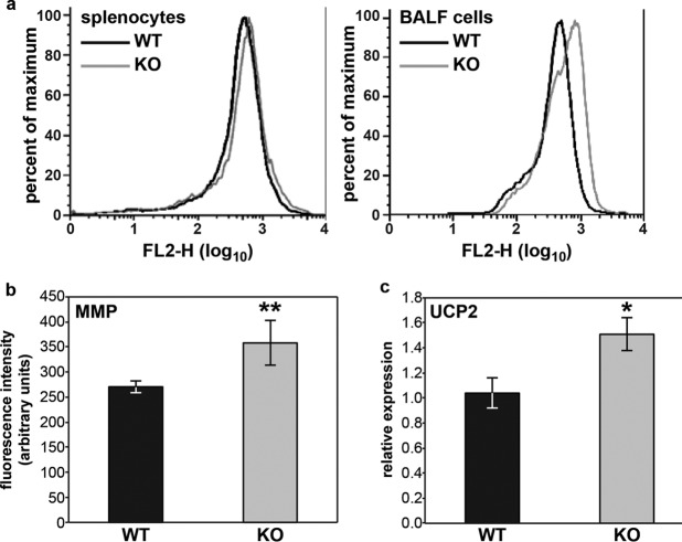
Fig 5.
Fus1−/− BALF cells show alterations of mitochondrial membrane potential (ΔΨ) and UCP2 expression after A. baumannii infection. (a) Evaluation of infection-induced changes of mitochondrial membrane potential (ΔΨ) in BALF cells and splenocytes from Fus1+/+ (WT) and Fus1−/− (KO) mice at 36 hpi. ΔΨ was measured via fluorescence-activated cell sorting analysis of cells stained with potential-dependent lipophilic cationic dye, JC-1, that is selectively accumulated in mitochondria, where it forms monomers at low ΔΨ or forms aggregates at high ΔΨ, which results in different fluorescence colors. Curves show right-shift (hyperpolarization) in FL-2 fluorescence observed in Fus1−/− cells (gray line) compared to wild-type cells (black line). (b) Expression of Ucp2 in BALF cells of infected mice at 36 hpi was assessed by real-time RT-PCR and normalized to GusB. All data represent means ± the standard errors. *, P < 0.05; **, P < 0.01 (Fus1+/+ versus Fus1−/−; n = 3 to 5 mice per group).

