Fig 1.
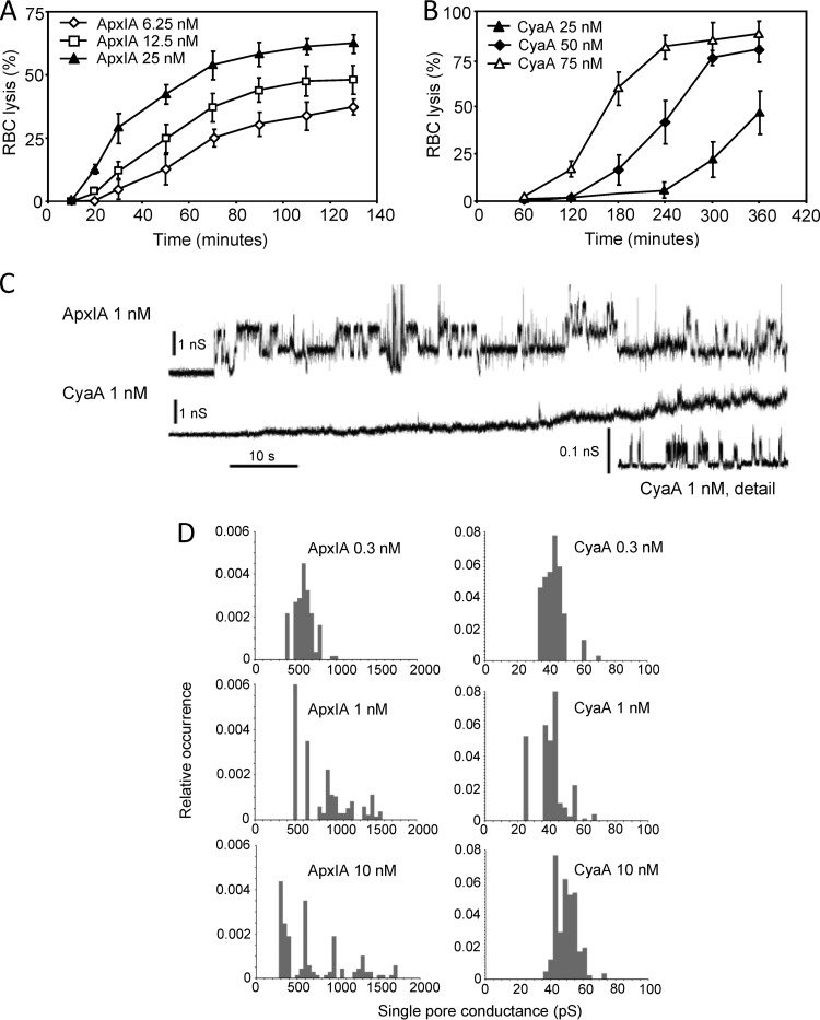
Sheep erythrocytes are susceptible to the pore-forming activity of ApxIA and CyaA in a concentration-dependent manner. Sheep erythrocytes (5 × 10⁸/ml) in HBSS buffer were incubated at 37°C in the presence of different concentrations of ApxIA (A) and CyaA (B). Hemolytic activity was measured as the amount of released hemoglobin by photometric determination (A541). The results represent average values from two independent experiments performed in duplicate. (C) Typical current traces of ApxIA and CyaA in asolectin lipid bilayers in 1 M KCl, 10 mM Tris, and 2 mM CaCl2 (pH 7.4). The ApxIA and CyaA concentration was 1 nM. Traces were recorded 60 s after ApxIA or CyaA addition. The applied membrane potential was 55 mV; the temperature was 25°C. All recordings were filtered at 100 Hz. The detailed recording of CyaA pores was performed at a higher current resolution. (D) The conductance of single ApxIA or CyaA pores was determined in 1 M KCl, 10 mM Tris, and 2 mM CaCl2 (pH 7.4) at membrane potentials of between 50 and 75 mV; the temperature was 25°C.

