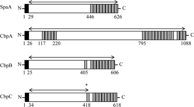
Fig 2.

Schematic representation of the domain structures of CBPs of E. rhusiopathiae Fujisawa. The signal sequence and choline-binding domain are indicated by black and gray boxes, respectively. Arrows indicate the regions of the recombinant proteins constructed. CbpC of Fujisawa is truncated by a frameshift mutation from a single nucleotide deletion (asterisk) in cbpC, and therefore, the schematic of CbpC is depicted as if it has no mutation. Numbers indicate the positions of amino acids.
