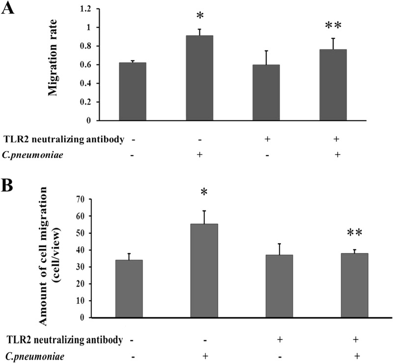
Fig 3.
Effects of TLR2 on rVSMC migration induced by C. pneumoniae infection. The TLR2-neutralizing antibody was added 1 h before C. pneumoniae infection. (A) Wound healing assay. “Scratch wounds” were created by scraping the confluent cell monolayer with a sterile pipette tip, and then cells were infected with C. pneumoniae at an infectious dose of 5 × 10⁵ IFU. Photographs were taken of the same wounded area of each well at 0 h and 24 h. The scratched regions were photographed under an inverted Nikon microscope (×100 magnification) at 24 h after C. pneumoniae infection. Migration velocity is presented as a ratio of the cellular recoverage area to the whole wound area. *, P < 0.05 versus control; **, P < 0.05 versus C. pneumoniae infection group. (B) Transwell migration assay. Cell morphology was observed by staining with 0.1% crystallin violet. The number of cells that had migrated through the pores was quantified by counting nine independent visual fields using a microscope (×200 magnification). *, P < 0.05 versus control; **, P < 0.05 versus the C. pneumoniae infection group.

