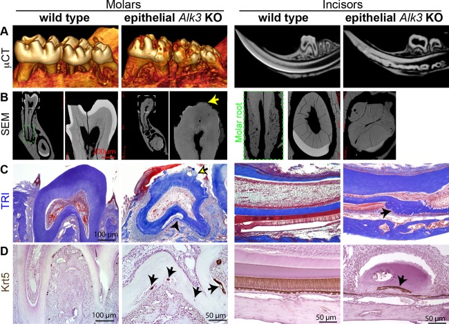
Fig 1.
Radiological and histological analyses of enamel defects and ectopic cellular cementum-like structures in AKO teeth. Krt5-rtTA/tetO-Cre/Alk3fl/fl (Alk3 KO [AKO]) mice and wild-type (WT) littermates were induced with Dox starting on E14.5. (A and B) Tooth samples were collected on P42 for μCT imaging (A) to examine molar surfaces or sagittal tooth sections or for backscatter SEM to show ultrastructures (B) with the green-outlined insert depicting the cellular cementum in a WT molar. (C and D) Sections of decalcified teeth were analyzed with TRI staining (C) or Krt5 IHC (D).

