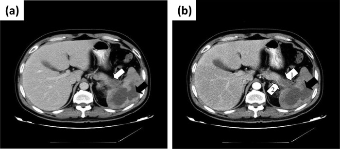
Fig 1.

(a) CT scan (portal phase) image of a hypodense area that invaded hilum of spleen, in the tail of pancreas (white arrow), and an irregular hypodense area with an unclear margin in the spleen (black arrow). (b) CT scan (arterial phase) image of nonenhancing hypodense area that invaded hilum of spleen, in the tail of pancreas (white arrow 1), and an irregular nonenhancing hypodense area in the spleen (black arrow). The splenic artery is slender (white arrow 2).
