
Figure 2.
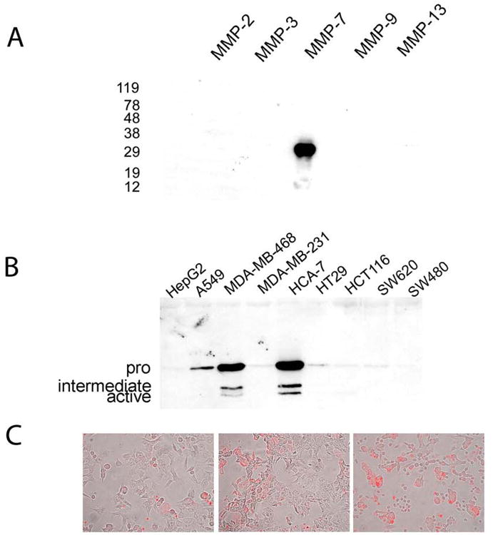
(a) Anti-MMP7 antibody specifically binds MMP-7. Western blotting of equimolar amounts (1 micro mole) of human recombinant MMP-2, MMP-3, MMP-7, MMP-9 and MMP-13 shows binding only to MMP-7. Sizes (kDa) of molecular weight markers are shown on the left. (b) MMP-7 expression in human adenocarcinoma cell lines. The cell lines used were HepG2 (hepatocellular carcinoma), A549 (lung adenocarcinoma), MDA-MB-468 (breast adenocarcinoma), MDA-MB-231 (breast adenocarcinoma), HCA-7 (colon adenocarcinoma), HT-29 (colon adenocarcinoma), HCT116 (colon adenocarcinoma), SW620 (lymph node metastasis of colon adenocarcinoma) and SW480 (colon adenocarcinoma). Bands representing the latent (30kDa), intermediate (approx 23kDa) and active (19kDa) forms of MMP-7 are evident in the strongest expressing samples MDA-MB-468 and HCA-7. Other positive samples show only the latent form. (c) Human colon adenocarcinoma cell lines display MMP-7 expression as detected by immunofluorescence (red fluorophore). Panels show from left to right HCT116, HCT-15 and SW620 cells stained for MMP-7 expression.