
Figure 3.
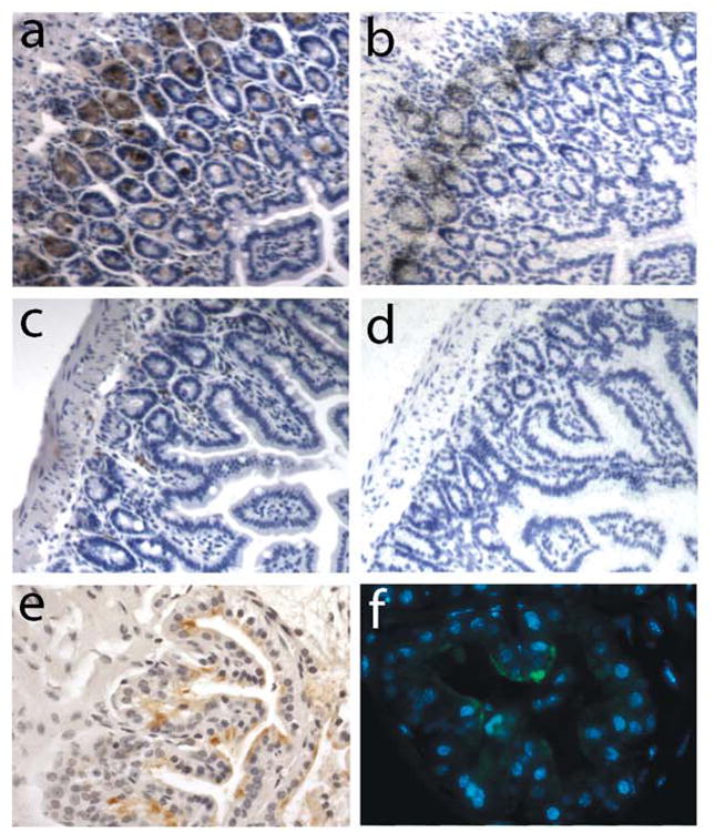
Expression of MMP-7 in murine tissue sections.(a,b) Section of small intestine from a wild-type C57bl/6 mouse showing immunohistochemical (a) and in situ hybridization (b) detection of MMP-7 expressed in the Paneth cells. (c, d) Sections of small intestine from a MMP-7-/- mouse demonstrating no signal by immunohistochemisty (c) or in situ hybridization (d) in Paneth cells. (e,f) Sections of ventral prostate from male C57bl/6 mice harvested 2-days post androgen ablation. Immunoperoxidase (e) and immunofluorescence (green fluorophore) (f) signals demonstrate localization of the induced MMP-7 protein to the luminal surface of the glands.