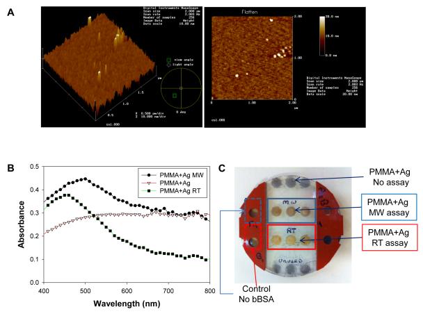
Figure 2.
Atomic force microscopy images of the PMMA platform coated with 2 nm silver nanoparticle film. (B) UV-Vis absorbance of blank PMMA and silver nanoparticle coated PMMA platform before and after the completion of the bioassay at room temperature (RT) and microwave heating (MW). (C) Real-color photograph of the silvered PMMA platform after the model bioassay was completed. The silicon isolator in the middle was removed to visually demonstrate the color of the wells.

