
Fig 5.
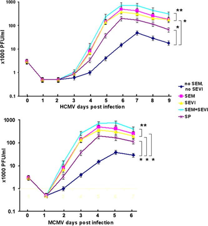
Viral growth curve assay. (Upper panel) HCMV infection of MRC-5 cells. MRC-5 cells were infected with virus alone or with virus treated with SP (1:1,000 dilution), SEM amyloids (5 μg/ml), SEVI (5 μg/ml), or SEM amyloids (5 μg/ml) plus SEVI (5 μg/ml) at an MOI of 0.1 for 24 h (as indicated). Each day for a period of 9 days, one well of each culture was collected (along with medium) and stored at −80°C. The collected cells (with medium) were freeze-thawed for three cycles and then centrifuged at 8,000 × g for 20 min to remove the cellular debris. To perform a PFU assay, a 25-μl supernatant of each sample was used to infect the MRC-5 cells in triplicates. (Lower panel) MCMV infection in NIH 3T3 cells. The procedure is the same as that used for HCMV infection of MRC-5 cells, except that the infection period was 6 days. A Student t test was used to statistically analyze the difference between the groups versus virus alone (*, P < 0.001) and that of the groups of SEM+SEVI versus SEM or SEVI (**, P < 0.005).