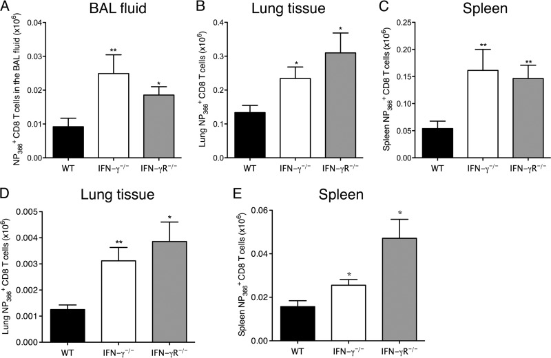
Fig 3.
Increased numbers of antigen-specific CD8 T cells in IFN-γ−/− and IFN-γR−/− mice after HKx31 infection. C57BL/6 WT, IFN-γ−/−, and IFN-γR−/− mice were infected with 50,000 PFU of influenza A/HK/x31 virus. NP366+ CD8+ T cell numbers were examined in the BAL fluid (A), lung tissue (B), and spleens (C) of the mice on day 28 p.i. and in the lung tissue (D) and spleens (E) on day 120 p.i. The numbers of CD8+ NP366+ cells (means ± SEM) shown were calculated from the total counts and the percentages of cells staining positive. *, P < 0.05; **, P < 0.01 (determined using Student's t test). Data are representative of at least 2 independent experiments with 4 or 5 mice per group.

