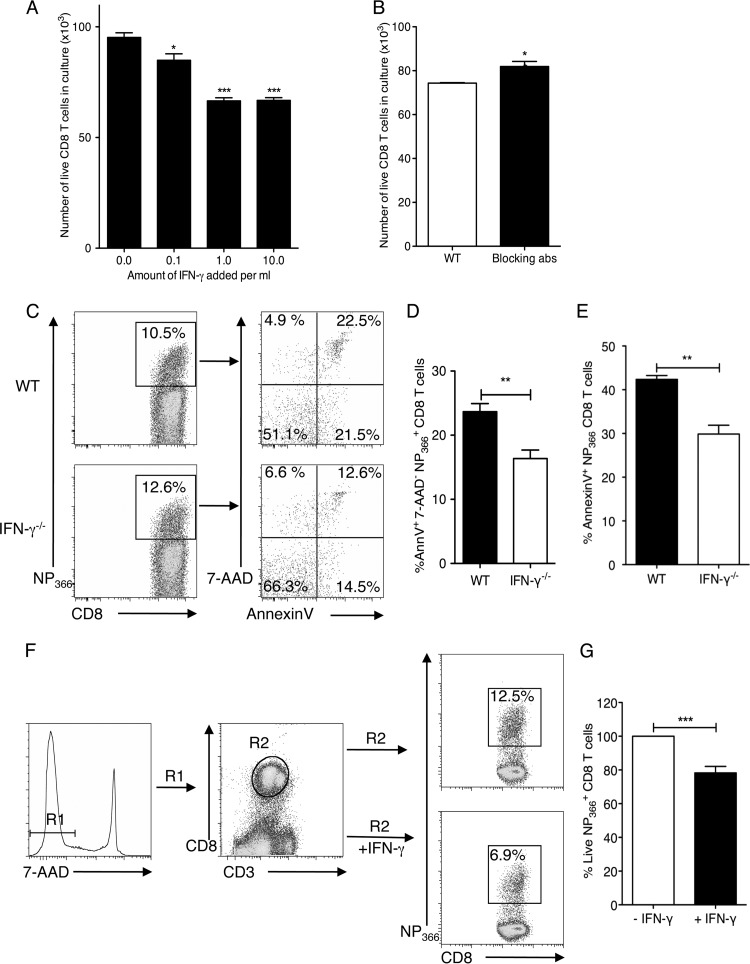
Fig 4.
The presence of IFN-γ leads to decreased CD8 T cell survival in culture and in influenza virus-specific CD8 T cells in the lungs of infected mice. CD8 T cells were purified from the spleens and lymph nodes of WT and IFN-γ−/− mice. IFN-γ−/− CD8 T cells were stimulated in vitro with phorbol myristate acetate (PMA) and ionomycin and cultured in the absence or presence of increasing amounts of recombinant IFN-γ in culture for 48 h. (A) Cells were stained with anti-CD3 and anti-CD8 antibodies and with 7-AAD. Live cells (7-AAD negative) were counted in all cultures and plotted. (B) WT CD8 T cells were stimulated in the presence or absence of blocking antibodies to IFN-γ and IFN-γR1 for 48 h. Live cells were counted and plotted after 48 h. (C) C57BL/6 WT or IFN-γ−/− mice were infected with 5 PFU of influenza A/PR/8/34 virus, and lungs were harvested on day 12 p.i. The CD8+ T cells from the lungs were isolated and stained with annexin V and 7-AAD. Representative flow cytometry plots of annexin V and 7-AAD staining of WT and IFN-γ−/− mice are shown. (D and E) The percentages of cells positive for annexin V and 7-AAD (D) or annexin V only (E) were plotted. (F) IFN-γ−/− mice were infected with 5 PFU of influenza A/PR/8/34 virus, and CD8+ T cells were harvested on day 14 p.i. The Ficoll-enriched cells were cultured in the presence or absence of rIFN-γ for 24 h. Live NP366+ CD8+ T cell proportions were examined in the culture according to the gating strategy. (G) The NP366+ CD8+ T cells in the cultures without IFN-γ were considered to represent 100%, and the percentage of live NP366+ CD8+ T cells in the cultures with IFN-γ were calculated accordingly. Means ± SEM are shown and were calculated from the percentages of cells staining positive. Data are representative of two or three independent experiments. *, P < 0.05; **, P < 0.01; ***, P < 0.001.

