Fig 1.
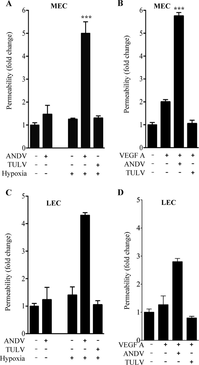
Hypoxia induces hyperpermeability of ANDV-Infected ECs. ECs were grown in CO2 (5%) incubators that regulate O2 levels by nitrogen displacement, resulting in normoxic (20% O2) or hypoxic (1% O2) conditions for cell growth. MECs (A and B) and LECs (C and D) were plated on vitronectin-coated Transwell inserts and grown under normoxic conditions (47, 49–52). Confluent EC monolayers were mock infected or infected with ANDV or TULV at an MOI of 0.5. Two days postinfection, ECs were growth factor starved overnight and incubated under normoxic conditions (control) or hypoxic conditions for 18 h (A and C). In parallel experiments, MECs and LECs (B and D) were stimulated with VEGF under normoxic conditions. Changes in monolayer permeability were assessed by adding FITC-dextran (40 kDa) to the upper chamber and monitoring its presence in lower chambers (A, B, C, and D) (47, 49–52). The data represent results from four independent experiments. Results are expressed as the fold increase in monolayer permeability over normoxic controls (**, P < 0.01; ***, P < 0.001).

