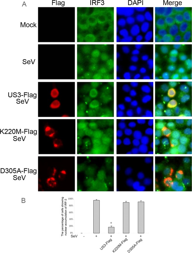
Fig 4.
US3 blocks SeV-induced IRF3 nuclear translocation. (A) HeLa cells were transfected with empty vector, Flag-tagged US3 WT, US3 K220M, or US3 D305A expression plasmid. Twenty-four hours posttransfection, cells were infected with 100 HAU/ml of SeV or mock-infected for 8 h as indicated. Cells were stained with mouse anti-Flag MAb and rabbit anti-IRF3 pAb. FITC-conjugated goat anti-rabbit (green) and TRITC-conjugated goat anti-mouse (red) were used as the secondary antibodies. Cell nuclei (blue) were stained with Hoechst 33258. The images were obtained by fluorescence microscopy using a 40× lens objective. (B) Statistical analysis of the percentage of cells exhibiting nuclear accumulation of IRF3 in panel A. Statistical analysis was performed using the Student t test. *, P < 0.05.

