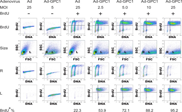
Fig 2.
Expression of GPC1 robustly stimulates DNA replication, resulting in an aneuploid DNA profile. U87-MG cells were infected with different doses of control or GPC1 adenovirus for 48 h as indicated and pulse-labeled with BrdU for 30 min at harvest and flow cytometry was performed after immunofluorescence staining for BrdU and propidium iodide (PI) staining for chromosomal DNA. Description of rows of panels: BrdU, analysis of PI labeling as a measure of DNA content (x axis) and BrdU incorporation as a measure of DNA synthesis (y axis); size, analysis of forward scatter (FSC) as a measure of nuclear size (x axis) and side scatter (SSC) as a measure of nuclear internal complexity (y axis). Flow cytometry with the FSC-SSC setting revealed that the input (nuclei) contained two populations of different size (R, regular size; L, enlarged size). R, analysis and axis designations as in the first row but gated to nuclei of regular size only. L, analysis and axis designations as in the first row but gated to nuclei of large size only. BrdU incorporation in the cells was dramatically induced by GPC1 transduction, and two cell populations with either regular or enlarged nuclei were formed following ectopic expression of GPC1. BrdU incorporation and aneuploidy occurred primarily in the cell population with large nuclei.

