
Fig 6.
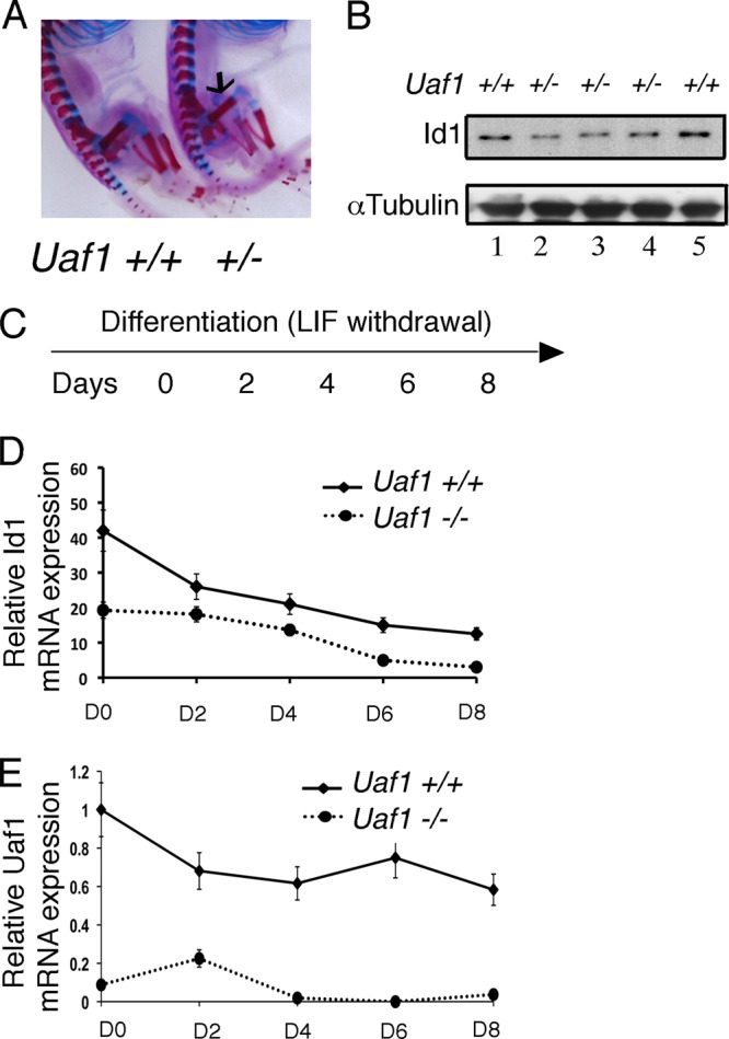
Uaf1 deficiency results in decreased Id1 expression. (A) Skeletal staining of Uaf1+/− and Uaf1+/+ E19 embryos. The arrow indicates the location of the femur. (B) Immunoblots of the lysates from femurs (in panel A) of Uaf1+/− and Uaf1+/+ E19 embryos. (C to E) Uaf1−/− and Uaf1+/+ mESCs were cultured without LIF in order to promote differentiation. RNA was then isolated at days (D) 0, 2, 4, 6, and 8 for quantification of Id1 and Uaf1 by qPCR. Id1 or Uaf1 expression upon differentiation of Uaf1−/− and Uaf1+/+ mESCs is shown. The data are shown as relative expression of Id1 or Uaf1 mRNA in Uaf1−/− mESCs compared to the wild-type controls. The samples were normalized using GAPDH expression.